Raji-APC-Null Cells
| Product | Unit size | Cat. code | Docs. | Qty. | Price | |
|---|---|---|---|---|---|---|
|
Raji-APC-Null Cells Control APC for anti-immune checkpoint cell-based assay |
Show product |
3-7 x 10e6 cells |
raji-apc-null
|
|
||
|
Raji-APC-Null vial Additional cell vial |
Show product |
3-7 x 10e6 cells |
raji-apc-null-av
|
Notification: Reference #raji-apc-null-av can only be ordered together with reference #raji-apc-null.

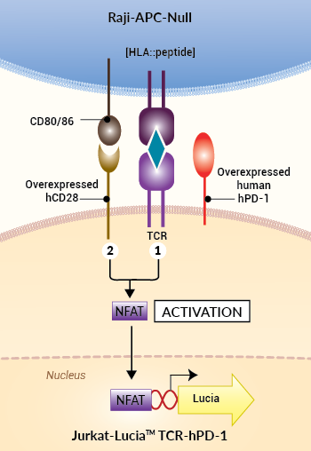
Principle of activating control APC for anti-immune checkpoint cell-based assay
 InvivoGen also offers:
• PD-1/PD-L1 BioIC™
• Anti-hPD-1 & hPD-L1 antibodies
• Anti-β-Gal antibodies
Activating control APC for anti-immune checkpoint cell-based assay
InvivoGen has developed Raji-APC-Null cells, as antigen-presenting cells (APCs), from the human B lymphoblastic Raji cell line.
• Raji-APC-Null cells – Antigen-Presenting Cells for Jurkat-Lucia™ TCR-derived cells
These cells differ from Raji-Null cells.
In addition to the high endogenous surface expression of CD80/86 activatory molecules, Raji-APC-Null cells feature the stable cell surface expression of a specific antigenic peptide complexed with HLA molecules ([HLA::peptide]). Thus, Raji-APC-Null, but not Raji-Null cells, can be used as activating APCs for Jurkat-Lucia™ TCR-derived cells.
A first signal (signal 1) is delivered upon recognition of [HLA::peptide] on Raji-APC cells by the TCR on Jurkat-Lucia™ cells. A second signal (signal 2), also known as the co-stimulation signal, is operated by the interaction of CD80/86 and CD28 molecules at the surface of the APC and Jurkat-Lucia™ TCR cells, respectively. Raji-APC-Null cells express immune checkpoints (ICs) at very low to barely detectable levels, so they serve as control APC for InvivoGen's anti-immune checkpoint cell-based assay (Bio-IC™).
Key features:
- Stable specific [HLA::peptide] expression
- Endogenous hCD80/86 expression
- Very low (barely detectable) expression levels to no expression of immune checkpoints (ICs)
Application:
Raji-APC-Null cells have been designed as control APCs for InvivoGen’s PD-1/PD-L1 Bio-IC™.
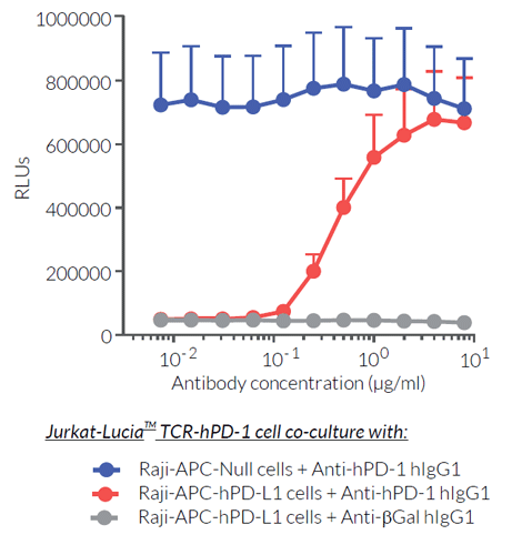
These cells do not provide an immune checkpoint signal to the Jurkat-Lucia™ TCR-hPD-1 effector reporter cells.
![]() Read our review on Immune Checkpoint Blockade
Read our review on Immune Checkpoint Blockade
![]() Learn more about Immune Checkpoint Antibodies.
Learn more about Immune Checkpoint Antibodies.
Specifications
Growth medium: IMDM, 2 mM L-glutamine, 25 mM HEPES, 10% (v/v) heat-inactivated fetal bovine serum (FBS), 100 U/ml penicillin, 100 µg/ml streptomycin, 100 µg/ml Normocin™
Antibiotic resistance: Blasticidin and G418 (Geneticin)
Quality Control:
- Activation of Jurkat-Lucia™ TCR-hPD-1 reporter cell line using Raji-APC-Null cells as antigen-presenting cells has been validated.
- The stability for 20 passages following thawing has been verified.
- This cell line is guaranteed mycoplasma-free.
Contents
- 3-7 x 10⁶ Raji-APC-Null cells in a cryovial or shipping flask
- 1 ml of Blasticidin (10 mg/ml)
- 1 ml of G418 (Geneticin) (100 mg/ml)
-
1 ml of Normocin™ (50 mg/ml). Normocin™ is a formulation of three antibiotics active against mycoplasmas, bacteria, and fungi.
![]() Shipped on dry ice (Europe, USA & Canada).
Shipped on dry ice (Europe, USA & Canada).
Details
The activation of T lymphocytes initiates their proliferation and yields a variety of effector functions that allow combating microbial infections, as well as developing tumors. The current paradigm is that full activation of T cells requires at least 2 signals upon contact with antigen-presenting cells (APCs) [1, 2].
Signal 1 is delivered through the interaction of the T cell receptor (TCR) and a specific antigenic peptide associated with an MHC (major histocompatibility complex) molecule on APCs. Signal 2 is delivered through the interaction of CD28, the prototypical T cell co-stimulatory molecule, and its ligands, CD80 or CD86, expressed by the APC. However, a number of other molecules, named immune checkpoints (IC), have been reported to regulate the onset and the limitations of T cell activities. PD-1 (programmed cell death 1) receptor and its ligand, PD-L1, are among the best characterized suppressive immune checkpoints [3].
Signal 1: TCR and [HLA::peptide]
The 'classical' and most represented TCR is an 80 to 90 kDa heterodimer composed of one α chain and one β chain. The αβTCR is a transmembrane protein expressed by developing and mature T cells. It features an extracellular ligand-binding pocket and a short cytoplasmic tail. Each αβTCR is restricted to a specific complex made of an antigenic peptide and a class I or class II MHC molecule. Human MHC molecules are also known as HLA (human leukocyte antigen). Because of its short cytoplasmic tail, the TCR, once engaged, lacks the ability to signal and requires non-covalent association with the CD3 to trigger downstream intracellular signaling and T cell activation [1, 2]. Importantly, signal 1 without co-stimulation results in T cell unresponsiveness or 'anergy', a tolerance mechanism that guards against premature activation.
Signal 2: CD28 and CD80/86
CD28 is a homodimeric and transmembrane protein expressed by T cells. Nearly all human CD4+ T cells and 50% of human CD8+ T cells express CD28. The CD28 interaction with CD80 (aka B7-1) or CD86 (aka B7-2) on APCs, in conjunction with TCR engagement, triggers a co-stimulation signal (signal 2). It results in T cell proliferation, cytokine production, cell survival, and cellular metabolism [1, 2].
IC signal: PD-1 and PD-L1
— PD-1 (programmed cell death 1; also known as CD279) is a type I transmembrane protein expressed at the cell surface of activated and exhausted conventional T cells. PD-1 is an inhibitory immune checkpoint that prevents T-cell overstimulation and host damage. PD-1 interaction with its ligands PD-L1 and PD-L2 induces inhibition of TCR signaling [3].
— PD-L1 (programmed cell death ligand 1; also known as CD274 or B7-H1) is a transmembrane protein expressed at the cell surface of hematopoietic and nonhematopoietic cells and is induced by pro-inflammatory cytokines, such as in the tumor microenvironment [3]. PD-L1 is one ligand for PD-1, an inhibitory immune checkpoint receptor that is expressed by activated and exhausted T cells. PD-1:PD-L1 interaction induces inhibition of TCR signaling, thereby preventing T-cell overstimulation and host damage [3].
References:
1. Budd R.C. & Fortner K.A., 2017. Chapter 12 - T Lymphocytes. Kelley and Firestein's Textbook of Rheumatology (Tenth Edition). pages 189-206.
2. Smith-Garvin J.E. et al., 2009. T Cell Activation. Ann. Rev. Immunol. 27:591-619.
3. Ribas A. and Wolchock J.D., 2018. Cancer immunotherapy using checkpoint blockade. Science. 359:1350-55.


