B16-Blue™ IFN-γ Cells
| Product | Unit size | Cat. code | Docs. | Qty. | Price | |
|---|---|---|---|---|---|---|
|
B16-Blue™ IFN-γ Cells Murine B16 melanoma IFN-γ reporter cells |
Show product |
3-7 x 10e6 cells |
bb-ifng
|
|
||
|
B16-Blue™ IFN-γ vial Additional cell vial |
Show product |
3-7 x 10e6 cells |
bb-ifng-av
|
Notification: Reference #bb-ifng-av can only be ordered together with reference #bb-ifng.
Murine Type II IFN Reporter Cells

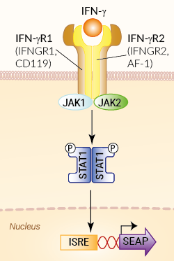
Signaling pathway in B16-Blue™ IFN-γ cells
B16-Blue™ IFN-γ cells were engineered from the murine B16 melanoma cell line to detect bioactive murine type II interferon IFN-γ by monitoring the activation of the JAK/STAT1 pathway. In addition, these cells can be used for screening antibodies or small molecule inhibitors targeting the IFN-γ pathway.
IFN-γ is a pleiotropic cytokine with anti-viral, anti-tumor, and immunomodulatory functions [1].
Cell line description
B16-Blue™ IFN-γ cells were generated by stable transfection with the secreted embryonic alkaline phosphatase (SEAP) reporter under the control of the ISG54 promoter. This promoter comprises IFN-stimulated response elements (ISRE) that are recognized by the ISGF3 complex. The binding of IFN-γ to its receptor triggers a signaling cascade leading to the activation of STAT1 and the subsequent production of SEAP. This can be readily assessed in the supernatant using QUANTI-Blue™ Solution, a SEAP detection reagent.
B16-Blue™ IFN-γ cells respond specifically to murine (m) IFN-γ and do not respond to human (h) IFN-γ. Of note they do not respond to mIFN-α/β (see figures).
Key features
- Fully functional murine IFN-γ signaling pathway
- Readily assessable STAT1-inducible SEAP reporter activity
- Strong response to murine IFN-γ
- No response to human IFN-γ
- No response to murine IFN-α/β (type I IFNs)
Applications
- Detection of murine IFN-γ
- Screening of anti-IFN-γ and anti-IFNGR antibodies
- Screening of small molecule inhibitors of the IFN-γ pathway
Reference:
1. Ivashkiv L.B., 2018. IFNγ: signalling, epigenetics and roles in immunity, metabolism, disease and cancer immunotherapy. Nat Rev Immunol. 18(9):545-558.
2. Platanias LC., 2005. Mechanisms of type-I- and type-II-interferon-mediated signalling. Nat Rev Immunol. 5(5):375-86.
Specifications
Detection range for murine IFN-γ: 0.1 ng - 1 µg/ml for mIFN-γ
Antibiotic resistance: Zeocin®
Quality control:
- B16-Blue™ IFN-γ cells were stimulated with murine IFN-α, IFN-β and IFN-γ. The cells produce SEAP only in response to murine IFN-γ
- These cells are guaranteed mycoplasma-free
Growth medium: DMEM, 10% (v/v) heat-inactivated fetal bovine serum, 2 mM L-glutamine, 100 µg/ml Normocin™, 100 U/ml penicillin, 100 µg/ml streptomycin
Back to the topContents
- 1 vial containing 3-7 x 10⁶ cells
- 1 ml of Zeocin® (100 mg/ml).
- 1 ml of Normocin™ (50 mg/ml).
- 1 ml of QB reagent and 1 ml of QB buffer (sufficient to prepare 100 ml of QUANTI-Blue™ Solution, a SEAP detection reagent)
![]() Shipped on dry ice (Europe, USA, Canada and some areas in Asia)
Shipped on dry ice (Europe, USA, Canada and some areas in Asia)
Details
Interferon-gamma (IFN-γ) is the sole member of the type II IFN family. It is secreted from CD4+ Th1 cells and activated NK cells. It plays a role in activating lymphocytes to enhance anti-microbial and anti-tumor effects [1-]. In addition, it plays a role in regulating the proliferation, differentiation, and response of lymphocyte subsets.
IFN-γ exerts its action by first binding to a heterodimeric receptor consisting of two chains, IFNGR1 and IFNGR2, causing its dimerization and the activation of specific Janus family kinases (JAK1 and JAK2) [4, 5]. Two STAT1 molecules then associate with this ligand-activated receptor complex and are activated by phosphorylation. Activated STAT1 molecules form homodimers and are translocated to the nucleus where they bind IFN-stimulated response elements (ISRE) in the promoter of IFN inducible genes.
1. Ivashkiv L.B., 2018. IFNγ: signalling, epigenetics and roles in immunity, metabolism, disease and cancer immunotherapy. Nat Rev Immunol. 18(9):545-558.
2. Shtrichman R. & Samuel CE., 2001. The role of gamma interferon in antimicrobial immunity. Curr Opin Microbiol. 4(3):251-9.
3. Sato A. et al., 2006. Antitumor activity of IFN-lambda in murine tumor models. J Immunol. 176(12):7686-94.
4. Platanias L.C., 2005. Mechanisms of type-I- and type-II-interferon-mediated signalling. Nat Rev Immunol. 5(5):375-86.
5. Schroder K. et al., 2004. Interferon-gamma: an overview of signals, mechanisms, and functions. J Leukoc Biol. 75(2):163-89.


