B16-Blue™ ISG-KO-STING Cells
| Product | Unit size | Cat. code | Docs. | Qty. | Price | |
|---|---|---|---|---|---|---|
|
B16-Blue™ ISG-KO-STING Cells Murine B16 melanoma - STING Knockout IRF-reporter cells |
Show product |
3-7 x 10e6 cells |
bb-kostg
|
|
||
|
B16-Blue™ ISG-KO-STING vial Additional cell vial |
Show product |
3-7 x 10e6 cells |
bb-kostg-av
|
Disclaimer: Reference #bb-kostg-av can only be ordered together with reference #bb-kostg.
STING Knockout IRF-Inducible SEAP Reporter B16 Melanocytes
B16-Blue™ ISG-KO-STING cells can be used for the study of the STING signaling pathway and for the detection of bioactive murine types I and II IFNs by monitoring the activation of the JAK/STAT pathway.
B16-Blue™ ISG-KO-STING cells were generated from the B16-Blue™ ISG cell line, a murine B16 melanoma-derived cell line, through stable knockout of the STING gene. They express the secreted embryonic alkaline phosphatase (SEAP) reporter gene under the control of the I-ISG54 promoter which is comprised of the IFN-inducible ISG54 promoter enhanced by a multimeric ISRE.
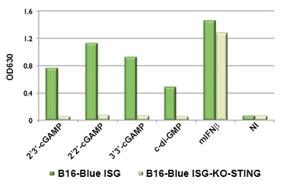
B16-Blue™ ISG-KO-STING cells do not respond to cytosolic DNA, DMXAA, canonical and non-canonical CDNs while retaining the ability to respond to type I and type II IFNs.
Stimulation of these cells with IFN triggers the activation of the I-ISG54 promoter and the production of SEAP. Levels of SEAP in the supernatant can be easily determined using QUANTI- Blue™, a reagent that turns purple/blue in the presence of SEAP, by reading the OD at 620-655 nm.
B16-Blue™ ISG-KO-STING cells are resistant to Zeocin®.
Back to the topSpecifications
Murine IFN Detection range
• mIFN-α: 10e2 - 10e4 IU/ml
• mIFN-β: 10e2 - 10e4 IU/ml
• mIFN-γ: 0.1 ng - 1 µg/ml
Antibiotic resistance: Zeocin®
Quality control:
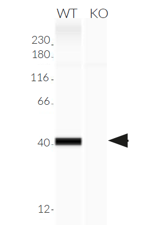
- STING knockout is verified by functional assays and DNA sequencing to confirm frameshift mutation/deletion
- These cells are guaranteed mycoplasma-free
Growth medium: DMEM, 10% (v/v) heat-inactivated fetal bovine serum, 2 mM L-glutamine, 100 µg/ml Normocin™, 100 U/ml penicillin, 100 µg/ml streptomycin
Back to the topContents
- 1 vial containing 3-7 x 10⁶ cells
- 1 ml of Zeocin® (100 mg/ml)
- 1 ml of Normocin™ (50 mg/ml)
- 1 ml of QB reagent and 1 ml of QB buffer (sufficient to prepare 100 ml of QUANTI-Blue™ Solution, a SEAP detection reagent)
![]() Shipped on dry ice (Europe, USA, Canada and some areas in Asia)
Shipped on dry ice (Europe, USA, Canada and some areas in Asia)


