Recombinant Human Fc-h4-1BBL Fusion Protein
| Product | Unit size | Cat. code | Docs. | Qty. | Price | |
|---|---|---|---|---|---|---|
|
Fc-h4-1BBL Soluble human 4-1BBL fused to an IgG1 Fc domain |
Show product |
50 µg |
fc-41bbl
|
|
Soluble human 4-1BBL (CD137L) fused to an IgG1 Fc domain

Potential applications of soluble Fc-h4-1BBL protein
Protein description
 InvivoGen also offers:
• Anti-IC cell-based assays (Bio-IC™)
• IC-expressing cell lines
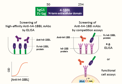
InvivoGen offers Fc-h4-1BBL, a soluble human 4-1BBL (CD137L) chimera protein generated by fusing the N-terminal extracellular domain of human 4-1BBL (aa 50-254) to the C-terminus of a human IgG1 Fc domain with a TEV (Tobacco Etch Virus) sequence linker.
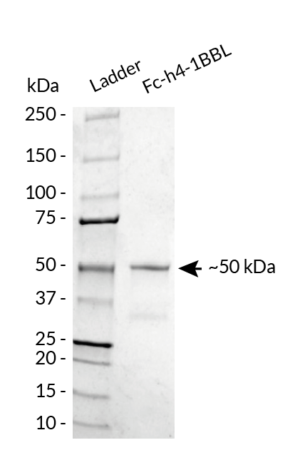
Fc-h4-1BBL has been produced in CHO cells and purified by affinity chromatography. It has an apparent molecular weight of ~50 kDa on an SDS‑PAGE gel.
4-1BBL background
4-1BBL (CD137L) is a Type II transmembrane protein and a member of the TNFR family known as the ligand for 4-1BB (CD137 or TNFSF9). 4-1BB is expressed on a multitude of cells including activated CD8+ and CD4+ T cells, and 4-1BBL is expressed on antigen-presenting cells (APCs) [1]. The 4-1BB/4-1BBL interaction triggers an NF‑κB‑dependent signaling, powerfully augmenting T cell activation, proliferation, and survival [1]. The 4-1BB/4-1BBL pair is thus considered a costimulatory immune checkpoint [1].
Applications
- Screening of high-affinity anti-human 4-1BBL monoclonal antibodies by ELISA
- Screening of anti-human 4-1BB monoclonal antibodies using competition assays
Quality control
- Size and purity confirmed by SDS-PAGE
- Protein validated by ELISA using an Anti-h4-1BBL monoclonal antibody
- Protein validated by flow cytometry using Jurkat-Lucia™ h4-1BB cells
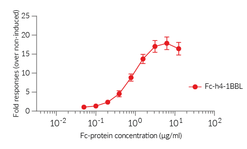
- Protein potency at triggering intracellular signaling validated using Jurkat-Lucia™ h4-1BB cells
Reference
1. Bartkowiak, T. & Curran, M.A. 2015. 4-1BB Agonists: Multi-Potent Potentiators of Tumor Immunity. Front Oncol 5, 117.
Back to the topSpecifications
Protein construction: N-terminal extracellular domain of human 4-1BBL (aa 50-254) with an N-terminal human IgG1 Fc tag
Accession sequence: NM_003811.3
Species: Human
Source: CHO cells
Tag: N-terminal human IgG1 Fc
Total protein size: 205 a.a (secreted form)
Molecular weight: ~50 kDa (SDS-PAGE)
Purification: Protein G affinity chromatography
Purity: >91% (SDS-PAGE)
Quality control:
- The protein is validated by ELISA upon incubation with an anti-h4-1BBL monoclonal antibody.
- The protein is validated by flow cytometry using Jurkat-Lucia™ h4-1BB cells.
- The potency of Fc-h4-1BBL at triggering intracellular signaling is validated using Jurkat-Lucia™ h4-1BB cells.
- The absence of bacterial contamination (e.g. lipoproteins and endotoxins) is confirmed using HEK-Blue™ TLR2 and HEK-Blue™ TLR4 cellular assays.
Contents
- 50 μg of lyophilized Fc-h4-1BBL protein
- 1.5 ml of endotoxin-free water
![]() The product is shipped at room temperature.
The product is shipped at room temperature.
![]() Lyophilized protein should be stored at -20 ̊C.
Lyophilized protein should be stored at -20 ̊C.
![]() Resuspended protein is stable for up to 1 month when stored at 4°C, and 1 year when stored at -20°C
Resuspended protein is stable for up to 1 month when stored at 4°C, and 1 year when stored at -20°C
Avoid repeated freeze-thaw cycles.
Back to the topDetails
The 4-1BB/4-1BBL immune checkpoint
The 4-1BB/4-1BBL axis plays an important role in immune regulation and is considered an immune checkpoint (IC).
4-1BB (aka CD137 or TNFSF9) is a member of the TNFR (tumor necrosis factor receptor) superfamily. In humans, 4-1BB is expressed on a multitude of cells of the hematopoietic lineage. It is found on the surface of activated CD8+ and CD4+ T cells, including regulatory T cells (Tregs). Additionally, its expression can be induced on Natural Killer (NK) cells, B cells, monocytes, and dendritic cells (DCs) upon activation [1,2].
4-1BBL (aka CD137L) is also a member of the TNFR family and is known as the sole ligand for 4-1BB. In humans, 4-1BBL is expressed in T cells, B cells, DCs and macrophages [1,2].
The interaction of 4-1BB on T cells with its ligand, 4-1BBL (CD137L), on antigen-presenting cells (APCs), triggers a TRAF1/TRAF2/NF‑κB‑dependent signaling which powerfully augments T-cell activation, proliferation, and survival [1]. This co-stimulatory axis also mediates the generation of memory T cells [1]
The 4-1BB/4-1BBL immune checkpoint in cancer
4-1BB expression in activated T cells has been an attractive target for cancer immunotherapy. Different strategies have been explored.
Two human agonistic monoclonal antibodies (mAbs) directed at 4-1BB, utomilumab (PF-05082566) and urelumab (BMS-663513), have entered phase I/II clinical trials. Whereas the IgG2 utomilumab is safe with relatively low efficacy, the IgG4 urelumab has a great antitumor efficacy but causes liver damage [2,3]. Utomilumab is being extensively tested in combination with IC inhibitory antibodies (e.g. Anti-CTLA-4 and Anti-PD-1) to treat different tumors, such as non-small cell lung cancer, kidney, head, and neck cancer [1,3]. Of note, agonistic 4-1BB mAbs may act on the depletion of Tregs through Fc-mediated effector functions [3]. A new generation of 4-1BB agonistic mAbs, other than utomilumab and urelumab, are being tested and are referenced in [2].
4-1BB-derived domains have been widely studied as costimulatory domains for genetically engineered T cells expressing chimeric antigen receptors (CART). The activation of the 4-1BB signaling in these CART cells promotes the formation of memory CART cells and long-term immune response against cancer cells [2,3].
4-1BB is expressed in various types of hematologic cancers and 4-1BBL in tumor cells, including carcinoma and colon cancers. However, the precise functions of 4-1BB/4-1BBL remain unclear in these malignancies [1, 2].
References:
1. Bartkowiak, T. & Curran, M.A. 2015. 4-1BB Agonists: Multi-Potent Potentiators of Tumor Immunity. Front Oncol 5, 117.
2. Singh, R. et al., 2024. 4-1BB immunotherapy: advances and hurdles. Experimental & Molecular Medicine. 56(1):32-39.
3. Mascarelli, D.E, et al., 2021. Boosting Antitumor Response by Costimulatory Strategies Driven to 4-1BB and OX40 T-cell Receptors. Front. Cell Dev. Biol. 10.3389/fcell.2021.692982.


