SARS-CoV-2 Spike S1 Proteins
| Product | Unit size | Cat. code | Docs. | Qty. | Price | |
|---|---|---|---|---|---|---|
|
Spike-S1-His SARS-CoV-2 Spike S1-His fusion protein |
Show product |
50 µg |
his-sars2-s1
|
|
||
|
Spike-S1-Fc SARS-CoV-2 Spike S1-Fc fusion protein |
Show product |
50 µg |
fc-sars2-s1
|
— |
||
|
Spike-S1-His (D614G) SARS-CoV-2 Spike (D614G) S1-His fusion protein |
Show product |
50 µg |
his-sars2-s1g
|
— |
||
|
Spike-S1-Fc (D614G) SARS-CoV-2 Spike (D614G) S1-Fc fusion protein |
Show product |
50 µg |
fc-sars2-s1g
|
— |
SARS-CoV-2 Spike S1 subunit with C-term His- or Fc-tag
Protein description
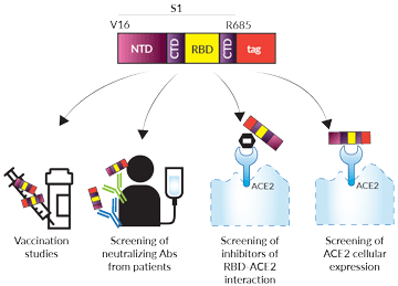
Potential applications of soluble tagged Spike S1 proteins
 InvivoGen also offers:
• Soluble Spike RBD proteins
• Soluble Nucleocapsid proteins
• Soluble Human ACE2 protein
The SARS-CoV-2 (2019-nCoV) Spike S1 subunit plays a crucial role in the viral entry into the target cell. The S1 subunit features an N-terminal domain (S1-NTD) and a C-terminal domain (S1-CTD). The S1-NTD is thought to mediate sugar-binding and the S1-CTD allows the virus to bind to ACE2 through the receptor-binding domain (RBD) [1-3]. In its resting conformation, S1 exerts a physical constraint on the Spike fusion subunit [3]. Early in the pandemic, the D614G amino acid mutation was identified within the Spike protein and has rapidly become the dominant variant around the world. The D614G mutation is located at the C-terminus of the S1 domain, near the furin cleavage site [4]. Research is ongoing to understand the exact mechanisms that drive conformation changes in S1 allowing subsequent membrane fusion events. S1 is a candidate for subunit vaccines against SARS-CoVs [5,6].
-
Spike-S1-His and Spike-S1-Fc were generated by fusing the SARS-CoV-2 Spike S1 [V16-R685] to a C-terminal poly-histidine sequence and human IgG1 Fc region, respectively. The S1 viral sequence used is from the original Wuhan-Hu-1 (D614) isolate.
-
Spike-S1-His (D614G) and Spike-S1-Fc (D614G) were generated by fusing the SARS-CoV-2 Spike S1 [V16-R685] to a C-terminal poly-histidine sequence and human IgG1 Fc region, respectively. The S1 viral sequence used is from the original Wuhan-Hu-1 isolate and contains the D614G mutation.
-
His- and -Fc-tagged proteins have been produced in HEK293 cells and CHO cells, respectively, and have been purified by affinity chromatography (See Specifications for more information).
Applications
- Vaccination studies: using combinations of Spike protein antigens and adjuvants
- Antibody screening: finding anti-Spike antibodies that can neutralize the SARS-CoV-2 infection
- Inhibitor screening: finding small molecules or antibodies able to block the SARS-CoV-2 RBD interaction with the ACE2 receptor
- ACE2 cellular expression screening: in primary isolated cells or transfected cells
Quality control
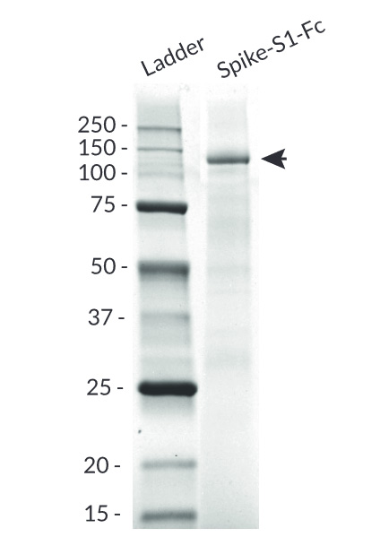
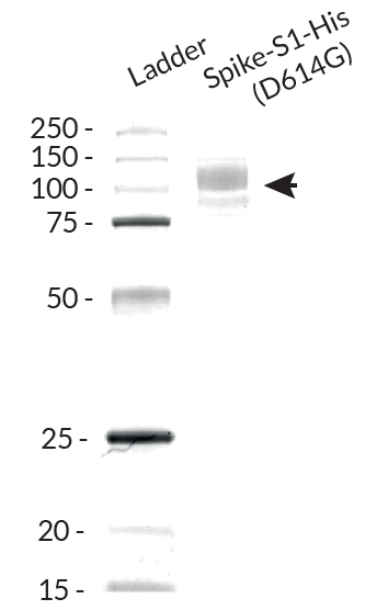
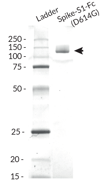
- Size and purity confirmed by SDS-PAGE
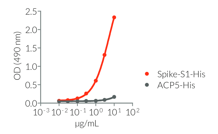
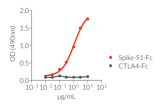
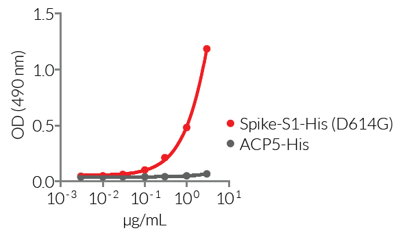
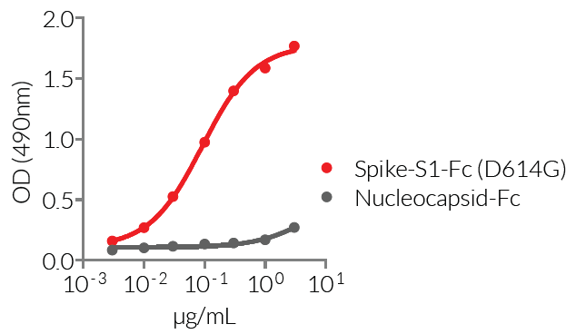
- Proteins validated by ELISA using a coated Anti-SARS-CoV-Spike-RBD hIgM mAb (clone CR3022)
![]() Read our reviews discussing SARS-CoV-2
Read our reviews discussing SARS-CoV-2
References
1. Li F., 2016. Structure, function, and evolution of coronavirus spike proteins. Annu.Rev. Virol. 3:237-261.
2. Li F. et al., 2005. Structure of SARS coronavirus spike receptor-binding domain complexed with receptor. Science. 309:1864-1868.
3. Walls A.C. et al.,2020. Structure, function, and antigenicity of the SARS-CoV-2 spike glycoprotein. Cell. 181(2):281-292.e6.
4. Korber B. et al., 2020. Tracking changes in SARS-CoV-2 Spike: evidence that D614G increases the infectivity of the COVID-19 virus. Cell. 182:1-16.
5. Wang N. et al., 2020. Subunit vaccines against emerging pathogenic human coronaviruses. Front. Microbiol. 11:298. DOI: 10.3389/fmicb.2020.00298.
6. Padron-Regalado E., 2020. Vaccines for SARS-CoV-2: Lessons from other coronavirus strains. Infect. Dis. Ther. DOI:10.1007/s40121-020-00300-x.
Specifications
Spike-S1-His
- Protein construction: S1 [V16-R685] from the Spike glycoprotein with a C-terminal poly-histidine tag
- Accession sequence: YP_009724390 (native sequence)
- Species: SARS-CoV-2 (2019-nCoV); Wuhan-Hu-1 (D614) isolate
- Tag: C-terminal poly-histidine (6 x His)
- Total protein size: 681 a.a. (secreted form)
- Molecular weight: ~ 123 kDa (SDS-PAGE gel)
- Purification: Ni2+ affinity chromatography
- Purity: >95% (SDS-PAGE)
-
Quality control:
- The protein has been validated by ELISA upon incubation with a coated Anti-SARS-CoV-Spike-RBD hIgM mAb (clone CR3022)
- The absence of bacterial contamination (e.g. lipoproteins and endotoxins) has been confirmed using HEK-Blue™ TLR2 and HEK-Blue™ TLR4 cellular assays.
Spike-S1-Fc
- Protein construction: S1 [V16-R685] from the Spike glycoprotein with a C-terminal human IgG1 Fc tag
- Accession sequence: YP_009724390 (native sequence)
- Species: SARS-CoV-2 (2019-nCoV); Wuhan-Hu-1 (D614) isolate
- Tag: C-terminal human IgG1 Fc
- Total protein size: 920 a.a. (secreted form)
- Molecular weight: ~124 kDa (SDS-PAGE)
- Purification: Protein A affinity chromatography
- Purity: >95% (SDS-PAGE)
-
Quality control:
- The protein has been validated by ELISA upon incubation with a coated Anti-SARS-CoV-Spike-RBD hIgM mAb (clone CR3022)
- The absence of bacterial contamination (e.g. lipoproteins and endotoxins) has been confirmed using HEK-Blue™ TLR2 and HEK-Blue™ TLR4 cellular assays.
Spike-S1-His (D614G)
- Protein construction: S1 [V16-R685] from the Spike glycoprotein with a C-terminal poly-histidine tag
- Accession sequence: YP_009724390 (native sequence)
- Species: SARS-CoV-2 (2019-nCoV); Wuhan-Hu-1 (D614) isolate with D614G variation
- Tag: C-terminal poly-histidine (6 x His)
- Total protein size: 681 a.a. (secreted form)
- Molecular weight: ~ 114 kDa (SDS-PAGE gel)
- Purification: Ni2+ affinity chromatography
- Purity: >95% (SDS-PAGE)
-
Quality control:
- The protein has been validated by ELISA upon incubation with a coated Anti-SARS-CoV-Spike-RBD hIgM mAb (clone CR3022)
- The absence of bacterial contamination (e.g. lipoproteins and endotoxins) has been confirmed using HEK-Blue™ TLR2 and HEK-Blue™ TLR4 cellular assays.
Spike-S1-Fc (D614G)
- Protein construction: S1 [V16-R685] from the Spike glycoprotein with a C-terminal human IgG1 Fc tag
- Accession sequence: YP_009724390 (native sequence)
- Species: SARS-CoV-2 (2019-nCoV); Wuhan-Hu-1 (D614) isolate with D614G variation
- Tag: C-terminal human IgG1 Fc
- Total protein size: 920 a.a. (secreted form)
- Molecular weight: ~126 kDa (SDS-PAGE)
- Purification: Protein A affinity chromatography
- Purity >95% (SDS-PAGE)
-
Quality control:
- The protein has been validated by ELISA upon incubation with a coated Anti-SARS-CoV-Spike-RBD hIgM mAb (clone CR3022)
- The absence of bacterial contamination (e.g. lipoproteins and endotoxins) has been confirmed using HEK-Blue™ TLR2 and HEK-Blue™ TLR4 cellular assays.
Contents
Spike-S1-His, Spike-S1-Fc, Spike-S1-His (D614G), and Spike-S1-Fc (D614G) contents:
(Note that each protein is sold separately)
- 50 μg of lyophilized protein
- 1.5 ml of endotoxin-free water
![]() The product is shipped at room temperature.
The product is shipped at room temperature.
![]() Lyophilized protein should be stored at -20 ̊C.
Lyophilized protein should be stored at -20 ̊C.
![]() Resuspended protein is stable up to 1 month when stored at 4°C, and 1 year when stored at -20°C
Resuspended protein is stable up to 1 month when stored at 4°C, and 1 year when stored at -20°C
Avoid repeated freeze-thaw cycles.
Back to the top


