RAW-Lucia™ ISG-KO-IRF1 Cells
| Product | Unit size | Cat. code | Docs. | Qty. | Price | |
|---|---|---|---|---|---|---|
|
RAW-Lucia™ ISG-KO-IRF1 Cells Murine RAW 264.7 macrophages - IRF1 Knockout IRF-Lucia Luciferase Reporter Cells |
Show product |
3-7 x 10e6 cells |
rawl-koirf1
|
|
||
|
RAW-Lucia™ ISG-KO-IRF1 vial Additional cell vial |
Show product |
3-7 x 10e6 cells |
rawl-koirf1-av
|
Notification: Reference #rawl-koirf1-av can only be ordered together with reference #rawl-koirf1.
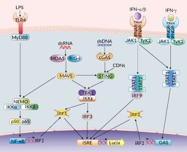
PRR signaling in RAW-Lucia™ ISG KO-IRF1 cells
IRF1 knockout reporter macrophages
RAW-Lucia™ ISG-KO-IRF1 cells were generated from the RAW-Lucia™ ISG cell line, which is derived from the RAW 264.7 murine macrophages, through the stable knockout of IRF1 gene.
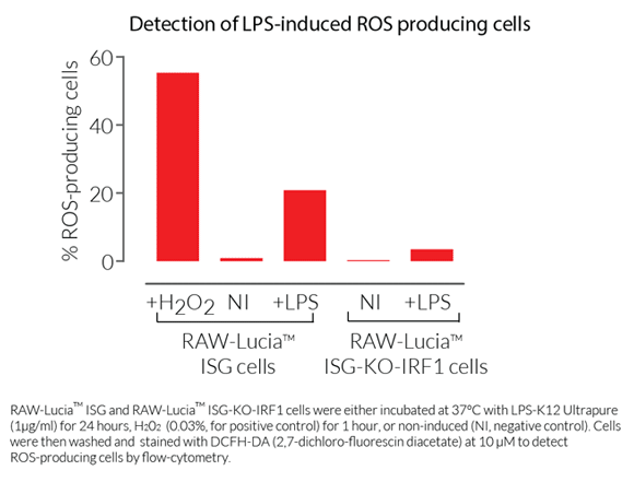
IRF1 (interferon regulatory factor 1) is the founding member of a family of nine transcription factors (IRF1 to IRF9) playing essential roles in immune responses. IRF1 is expressed at low levels in the cytoplasm of resting cells and is highly induced upon viral infections and inflammatory cytokine signaling. IRF1 promotes the expression of distinct sets of IFN-inducible genes (ISGs) depending on the cell type [1,2]. Notably, IRF1-induced expression upon TLR4 and IFN (mostly IFN-γ) signaling makes it a critical contributor to prolonged waves of ISG expression [2]. IRF1 is also involved in LPS-induced oxidative stress outcomes in macrophages, thus contributing to endotoxemia [3]. Of note, it has been proposed that LPS-mediated IRF1 expression in macrophages controls the production of oxidized mitochondrial DNA, and thus participates in the NLRP3 inflammasome activation [4].
The various biological functions in which IRF1 is involved support the search for IRF1-targeting therapeutic strategies [1-3,5]. Indeed, IRF1 is implicated in many inflammatory diseases, including inflammation-related cancers [2,5,6]. Unlike IRF3, IRF5, and IRF7 which are the principal mediators of IFN induction upon specific PRRs activation, IRF1 rather acts as an amplifier of the gene expression prompted by IRF3, IRF5, or IRF7 [7]. Thus, there is no clear phenotype for IRF1-deficient cells.
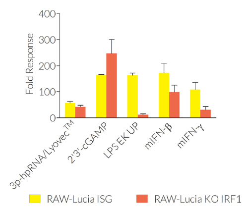
RAW-Lucia™ ISG-KO-IRF1 and RAW-Lucia™ ISG cells feature a Lucia luciferase reporter gene under the control of an ISG54 promoter enhanced by multimeric ISREs. Thus, activation of the IRF pathways can be readily assessed by monitoring the activity of the secreted Lucia luciferase in the supernatant using the QUANTI-Luc™ 4 Lucia/Gaussia detection reagent. Because IRF1 and other IRF-family members bind to closely related and overlapping ISRE motifs in the ISG54 promoter, RAW-Lucia™ ISG-KO-IRF1 cells display similar induction of the Lucia luciferase reporter when compared to their parental cells. Interestingly, however, the response of RAW-Lucia™ ISG-KO-IRF1 cells to lipopolysaccharide (LPS) is significantly altered when compared to RAW-Lucia™ ISG cells. The reporter activity in the RAW-Lucia™ ISG-KO-IRF1 cells provides a control of IRF induction (including IRF3, IRF5, IRF7 and IRF9) while testing IRF1-dependent phenotypes.
RAW-Lucia™ ISG-KO-IRF1 cells are resistant to Zeocin®.
Features of RAW-Lucia™ ISG KO-IRF1 cells:
- Biallelic knockout of the IRF1 gene
- Readily assessable Lucia luciferase activity
Applications for RAW-Lucia™ ISG KO-IRF1 cells:
- Defining the role of IRF1 in PRR-induced signaling
- Control of IRF (other than IRF1) induction while testing IRF1-dependent phenotypes (see Details)
Validation of RAW-Lucia™ ISG KO-IRF1 cells:
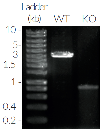
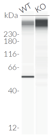
- IRF1 knockout verified by PCR, DNA sequencing, and Western blot
- Functionally tested
References:
1. Taniguchi T. et al., 2001. IRF family of transcription factors as regulators of host defense. Annual Rev. Immunol. 19:623.
2. Antonczyk A. et al., 2019. Direct inhibition of IRF-dependent transcriptional regulatory mechanisms associated with disease. Front. Immunol. 10:1176.
3. Deng S-Y., 2017. Role of interferon regulatory factor-1 in lipopolysaccharide-induced mitochondrial damage and oxidative stress responses in macrophages. Int. J. Mol. Med. 40:1261.
4. Zhong Z. et al., 2018. New mitochondrial DNA synthesis enables NLRP3 inflammasome activation. Nature. 560:198.
5. Thompson C.D. et al., 2018. Therapeutic targeting of IRFs: pathways-dependence or structure-based? Front. Immunol. 9:2622.
6. Hartley G. et al., 2017. Regulation of PD-L1 expression on murine tumor-associated monocytes and macrophages by locally produced TNF-α. Cancer Immunol. Immunother. 66:523.
7. Jeffries C.A., 2019. Regulating IRFs in IFN driven disease. Front. Immunol. 10: art325.
Specifications
Antibiotic resistance: Zeocin®
Growth Medium: DMEM, 4.5 g/l glucose, 2 mM L-glutamine, 10% (v/v) heat-inactivated fetal bovine serum, 100 U/ml penicillin, 100 μg/ml streptomycin, 100 μg/ml Normocin™ supplemented with Zeocin®.
Quality Control:
- IRF1 knockout is verified by PCR, DNA sequencing, Western blot, and functional assays.
- The stability for 20 passages, following thawing, has been verified.
- The cells are guaranteed mycoplasma-free.
Contents
- 1 vial containing 3-7 x 10⁶ cells
- 1 ml of Zeocin® (100 mg/ml)
- 1 ml of Normocin™ (50 mg/ml)
- 1 tube of QUANTI-Luc™ 4 Reagent, a Lucia luciferase detection reagent (sufficient to prepare 25 ml)
![]() Shipped on dry ice (Europe, USA, Canada and some areas in Asia)
Shipped on dry ice (Europe, USA, Canada and some areas in Asia)
Details
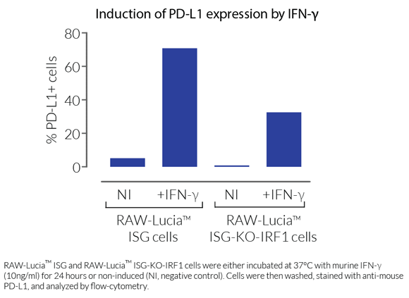
The reporter activity in the RAW-Lucia™ ISG-KO-IRF1 cells provides a control of IRF induction while testing IRF1-dependent phenotypes, such as LPS-induced ROS (reactive oxygen species) production [1], and inflammation-induced PD-L1 (programmed cell death ligand 1) expression [2].
1. Deng S-Y., 2017. Role of interferon regulatory factor-1 in lipopolysaccharide-induced mitochondrial damage and oxidative stress responses in macrophages. Int. J. Mol. Med. 40:1261.
2. Hartley G. et al., 2017. Regulation of PD-L1 expression on murine tumor-associated monocytes and macrophages by locally produced TNF-α. Cancer Immunol. Immunother. 66:523.


