


G-CSF Reporter HEK 293 Cells
| Product | Unit size | Cat. code | Docs. | Qty. | Price | |
|---|---|---|---|---|---|---|
|
HEK-Blue™ G-CSF Cells Human & Mouse G-CSF Reporter Cells |
Show product |
3-7 x 10e6 cells |
hkb-gcsf
|
|
||
|
HEK-Blue™ G-CSF vial Additional cell vial |
Show product |
3-7 x 10e6 cells |
hkb-gcsf-av
|
Notification: Reference #hkb-gcsf-av can only be ordered together with reference #hkb-gcsf.
Granulocyte Colony-Stimulating Factor Reporter Cells

Signaling pathway in HEK-Blue™ G-CSF cells
HEK-Blue™ G-CSF cells were engineered from the human embryonic kidney HEK 293 cell line to detect bioactive granulocyte colony-stimulating factor (G-CSF) by monitoring the activation of the JAK/STAT3 pathway. They can also be used for screening antibodies or small molecule inhibitors targeting the G-CSF pathway.
G-CSF is a secreted cytokine and hematopoietic growth factor. It regulates the differentiation, proliferation, and function of neutrophils [1].
Cell line description
HEK-Blue™ G-CSF cells were generated by stable transfection with the genes encoding for the human CSF receptor (CSFR, aka CD114), human STAT3c, and a STAT3-inducible secreted embryonic alkaline phosphatase (SEAP) reporter. The binding of G-CSF to its receptor triggers a signaling cascade leading to the activation of STAT3 and the subsequent production of SEAP. This can be readily assessed in the supernatant using QUANTI-Blue™ Solution, a SEAP detection reagent.
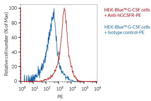
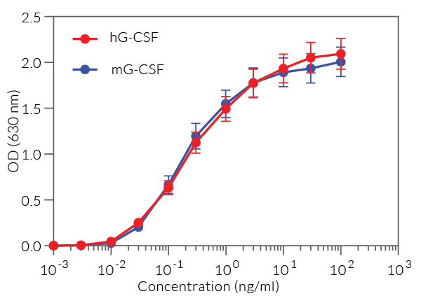
HEK-Blue™ G-CSF cells detect human and murine G-CSF (see figures). Of note, these cells also respond, to a weaker extent, to two other human STAT3-signaling cytokines, IL-6 and IL-27, as they endogenously express the gp130 co-receptor shared by these cytokine receptors. However, they do not respond to human type I IFNs (IFN-α/IFN-β) (see figures).
Key features
- Fully functional G-CSF signaling pathway
- Readily assessable STAT3-inducible SEAP reporter activity
- Strong response to human (h) G-CSF and mouse (m) C-CSF
- Weak response to IL-6 and IL-27
- No response to human type I IFNs
Applications
- Detection and quantification of human and murine G-CSF activity
- Screening of anti-G-CSF and anti-GCSFR antibodies
- Screening of small molecule inhibitors of the G-CSF pathway
Reference:
1. Martin KR, et al., 2021. G-CSF — A double-edged sword in neutrophil-mediated immunity. Sem. Immunol. 54:101516.
Figures


Specifications
Antibiotic resistance: Blasticidin, Hygromycin B, and Zeocin®
Growth medium: DMEM, 4.5 g/l glucose, 2-4 mM L-glutamine, 10% (v/v) fetal bovine serum, 100 U/ml penicillin, 100 μg/ml streptomycin, 100 μg/ml Normocin®
Specificity: Detects human and mouse G-CSF
Detection range:
- Detection range for human G-CSF: 300 pg/ml - 100 ng/ml
- Detection range for murine G-CSF: 300 pg/ml - 100 ng/ml
Quality Control:
- SEAP reporter activity in response toG-CSF is validated using functional assays.
- The expression of human GCSFR is confirmed by flow cytometry.
- The stability for 20 passages following thawing is confirmed.
- These cells are tested for mycoplasma contamination.
Contents
- 1 vial containing 3-7 x 10⁶ cells
- 1 ml Normocin® (50 mg/ml)
- 2 x 1 ml of HEK-Blue Selection (250X)
- 1 ml of QB reagent and 1 ml of QB buffer (sufficient to prepare 100 ml of QUANTI-Blue™ Solution, a SEAP detection reagent).
![]() Shipped on dry ice (Europe, USA, Canada)
Shipped on dry ice (Europe, USA, Canada)
Details
The granulocyte colony-stimulating factor (G-CSF) is a secreted cytokine and hematopoietic growth factor that belongs to the Type I/II cytokine receptor family. G-CSF regulates the differentiation, proliferation, and function of neutrophils [1]. It exerts its biological functions through the formation of a tetramer consisting of two G-CSF molecules and the homodimeric G-CSF receptor (GCSFR, aka CD114). The binding of G-CSF to its receptor triggers activation of the JAK/STAT signaling pathway [1,2]. STAT1, STAT3, and STAT5 are involved in this activation step and the induction of cellular proliferation [1,2]. Studies in humans and mice indicate that while G-CSF contributes to host defense against pathogens, it can also have detrimental effects by promoting inflammatory diseases and cancer [1,2].
References:
1. Martin KR., et al., 2021. G-CSF — A double-edged sword in neutrophil-mediated immunity. Sem Immunol. 54:101516.
2. Park, DS., et al., 2022. A review of granulocyte colony-stimulating factor receptor signaling and regulation with implications for cancer. Front Oncol. DOI: 10.3389/fonc.2022.932608.