RANKL Reporter HEK 293 Cells
| Product | Unit size | Cat. code | Docs. | Qty. | Price | |
|---|---|---|---|---|---|---|
|
HEK-Blue™ RANKL Cells Human & Mouse RANKL Reporter Cells |
Show product |
3-7 x 10e6 cells |
hkb-rankl
|
|
||
|
HEK-Blue™ RANKL vial Additional cell vial |
Show product |
3-7 x 10e6 cells |
hkb-rankl-av
|
Notification: Reference #hkb-rankl-av can only be ordered together with reference #hkb-rankl.
RANKL Reporter Cells

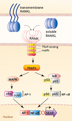
Signaling pathways in HEK-Blue™ RANKL cells
HEK-Blue™ RANKL cells were engineered from the human embryonic kidney HEK293 cell line to detect bioactive human and murine RANKL (Receptor Activator of NF-κB Ligand) by monitoring the activation of the NF-κB and AP-1 pathways. In addition, these cells can be used for screening antibodies or small molecule inhibitors targeting the RANKL pathway.
RANKL is a member of the TNF (Tumor Necrosis Factor) superfamily. This cytokine exists as a soluble or transmembrane protein produced by osteoblasts and activated T cells [1]. RANKL binding to its receptor RANK and the subsequent signaling events play a pivotal role in bone remodeling and dendritic cell survival, thereby enhancing the induction of T cell responses [1].
Cell line description
HEK-Blue™ RANKL cells were generated by stable transfection with the gene encoding for human RANK (with all three functional TRAF-binding motifs) and an NF-κB/AP1-inducible secreted embryonic alkaline phosphatase (SEAP) reporter. The binding of RANKL to its receptor triggers a signaling cascade leading to the activation of NF-κB/AP1, and the subsequent production of SEAP. This can be readily assessed in the supernatant using QUANTI-Blue™ Solution, a SEAP detection reagent.
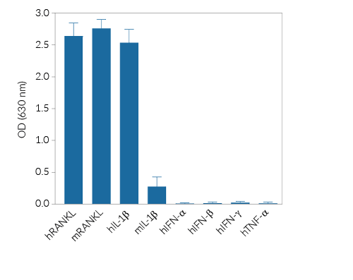
HEK-Blue™ RANKL cells respond to human (h) and mouse (m) RANKL (see figures). Of note, these cells also respond to hIL-1β and, to a weaker extent, mIL-1β, as they endogenously express the IL-1β receptor. They are not responsive to human IFN-α, IFN-β, IFN-γ, and TNF-α. They can also be used for screening and release assay of molecules that inhibit RANKL signaling, such as denosumab, a monoclonal antibody targeting RANKL (see figures).
Key features
- Fully functional RANKL signaling pathway
- Readily assessable NF-κB/AP1-inducible SEAP reporter activity
- Strong response to human (h) and murine (m) RANKL
- Response to hIL-1β and, to a weaker extent, mIL-1β
- No response to hIFN-α, hIFN-β, hIFN-γ, and hTNF-α
Applications
- Detection and quantification of human and murine RANKL activity
- Screening of anti-RANKL and anti-RANK antibodies
- Screening of small molecule inhibitors of the RANKL pathway
References:
1. Cheng ML. & Fong L., 2014. Effects of RANKL-targeted therapy in immunity and cancer. Front. Oncol. 3:329.
2. Ahern E.et al., 2018. Roles of the RANKL-RANK axis in anti-tumour immunity — implications for therapy. Nat. Rev. Clin. Oncol. 15:676-93.
3. Nakai Y.et al., 2019. Efficacy of an orally active small-molecule inhibitor of RANKL in bone metastasis. Bone Res. 7:1.
Specifications
Antibiotic resistance: Blasticidin, Zeocin®
Growth medium: DMEM, 4.5 g/l glucose, 2-4 mM L-glutamine, 10% (v/v) heat-inactivated fetal bovine serum, 100 U/ml penicillin, 100 μg/ml streptomycin, 100 μg/ml Normocin™
Guaranteed mycoplasma-free
Specificity: human and mouse RANKL
Detection range:
- 3 - 100 ng/ml for human RANKL
- 1 - 100 ng/ml for murine RANKL
Contents
- 1 vial containing 3-7 x 10⁶ cells
- 1 ml of Blasticidin (10 mg/ml)
- 1 ml of Zeocin® (100 mg/ml)
- 1 ml of Normocin™ (50 mg/ml)
- 1 ml of QB reagent and 1 ml of QB buffer (sufficient to prepare 100 ml of QUANTI-Blue™ Solution, a SEAP detection reagent)
![]() Shipped on dry ice (Europe, USA, Canada and some areas in Asia)
Shipped on dry ice (Europe, USA, Canada and some areas in Asia)
Details
Receptor Activator of NF-κB Ligand (RANKL), also known as member 11 of the tumor necrosis factor (TNF) superfamily (TNFSF11) or TNF-related activation-induced cytokine (TRANCE), exists as a transmembrane or soluble protein produced by osteoblasts and activated T cells [1]. RANKL binds to its receptor RANK via an obligate trimer configuration [1, 2].
RANKL/RANK signaling plays a pivotal role in bone remodeling and dendritic cell survival, thereby enhancing the induction of T cell responses [1]. Upon RANKL binding, RANK trimers recruit TNF receptor-associated factor (TRAF) adaptor proteins, such as TRAF6, to TRAF-binding motifs within their cytoplasmic domains [1].
The TRAF6 signaling cascade results in the activation of NF-κB and AP-1 transcription factors. Multiple efforts have focused on the development of anti‑RANKL antibodies or small‑molecule inhibitors for blocking RANKL/RANK signaling to reduce osteoporosis, prevent skeletal-related events (SREs) from bone metastasis in cancer, or improve anti-tumor immunity [1-3].
1. Cheng ML. & Fong L., 2014. Effects of RANKL-targeted therapy in immunity and cancer. Front. Oncol. 3:329.
2. Ahern E. et al., 2018. Roles of the RANKL-RANK axis in anti-tumour immunity — implications for therapy. Nat. Rev. Clin. Oncol. 15:676-93.
3. Nakai Y. et al., 2019. Efficacy of an orally active small-molecule inhibitor of RANKL in bone metastasis. Bone Res. 7:1.


