


IL-36 Reporter HEK 293 Cells
| Product | Unit size | Cat. code | Docs. | Qty. | Price | |
|---|---|---|---|---|---|---|
|
HEK-Blue™ IL-36 Cells Human IL-36 Reporter Cells |
Show product |
3-7 x 10e6 cells |
hkb-hil36r
|
|
||
|
HEK-Blue™ IL-36 vial Additional cell vial |
Show product |
3-7 x 10e6 cells |
hkb-hil36r-av
|
Interleukin-36 Reporter Cells

Signaling pathway in HEK-Blue™ IL-36 cells
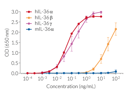
HEK-Blue™ IL-36 cells were engineered from the human embryonic kidney HEK 293 cell line to detect bioactive interleukin-36 (IL-36) by monitoring the activation of NF-κB/AP-1 pathways. Three isoforms, IL-36α, IL-36β, and IL-36γ, mediate pro-inflammatory functions, while a fourth one, IL-36Ra, acts as an antagonist [1,2].
Key features:
- Fully functional IL-36 signaling pathway
- Readily assessable NF-κB/AP-1-inducible SEAP reporter activity
- Strong response to human (h) IL-36α and IL-36γ
- Moderate response to hIL-36β
- No response to mouse (m) IL-36α
Applications:
- Detection and quantification of hIL-36 activity
- Screening of anti-IL-36 and anti-IL-36 receptor antibodies
- Screening of small molecule inhibitors of the IL-36 pathway
References:
1. Buhl A-L. & Wenzel J., 2019. Interleukin-36 in infectious and inflammatory skin diseases. Front Immunol. 10:1162.
2. Zhou L. & Todorovic V., 2021. Interleukin-36: Structure, Signaling and Function. Adv Exp Med Biol. 21:191-210.
Figures


Specifications
Cell type: Epithelial
Tissue origin: Human Embryonic Kidney
Target: IL-36α, IL-36β, IL-36γ
Specificity: Human
Reporter gene: SEAP
Antibiotic resistance: Blasticidin and Zeocin®
Detection range: 30 pg/ml - 100 ng/ml (hIL-36α and hIL-36γ), 10 ng/ml - 100 ng/ml (IL-36β)
Growth medium: Complete DMEM (see TDS)
Growth properties: Adherent
Mycoplasma-free: Verified using Plasmotest™
Quality control: Each lot is functionally tested and validated.
Back to the topContents
HEK-Blue™ IL-36 Cells (hkb-il36r)
- 1 vial containing 3-7 x 10⁶ cells
- 1 ml Normocin® (50 mg/ml)
- 1 ml Blasticidin™ (10 mg/ml)
- 1 ml Zeocin® (100 mg/ml)
-
1 ml of QB reagent and 1 ml of QB buffer (sufficient to prepare 100 ml of QUANTI-Blue™ Solution, a SEAP detection reagent)
HEK-Blue™ IL-36 vial (hkb-il36r-av)
- 1 vial containing 3-7 x 10⁶ cells
![]() Shipped on dry ice (Europe, USA, Canada, and some areas in Asia)
Shipped on dry ice (Europe, USA, Canada, and some areas in Asia)
Notification: Reference #hkb-il36r-av can only be ordered together with reference #hkb-il36r.
Back to the topDetails
Cell line description
HEK-Blue™ IL-36 cells were generated by stable transfection with the genes encoding for the human IL-36 heterodimeric receptor, comprising the IL-1R6 (aka IL-36R) and IL-1R3 (aka IL-1RAcP) subunits, and an NF-κB/AP-1-inducible secreted embryonic alkaline phosphatase (SEAP) reporter. The binding of IL-36 to its receptor triggers a signaling cascade leading to the NF-κB/AP-1 activation and the subsequent production of SEAP.
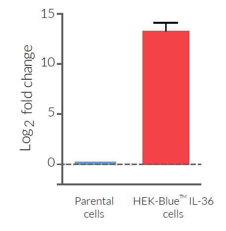
HEK-Blue™ IL-36 cells respond to human (h) but not murine (m) IL-36 agonist isoforms. Of note, they are more sensitive to hIL-36α and hIL-36γ compared to hIL-36β (see figures). HEK-Blue™ IL-36 cells maintain their responses to other cytokines that signal through NF-κB/AP-1, such as hIL-1β and hTNF-α (see figures).
IL-36 background
The cytokine interleukin 36 (IL-36) belongs to the IL-1 superfamily. Three isoforms, IL-36α, IL-36β, and IL-36γ, mediate pro-inflammatory functions, while a fourth one, IL-36Ra, acts as an antagonist [1,2]. IL-36 signalization requires the formation of a complex comprised of two subunits, IL-1R6 (aka IL-36R) and IL-1R3 (aka IL-1RAcP, IL-1 receptor accessory protein). The binding of agonist ligands to the IL-36R allows the recruitment of IL-1RAcP and the production of pro-inflammatory cytokines and chemokines through the activation of NF-κB and AP-1 [1,2]. The IL-36Ra antagonist inhibits the signaling by binding to IL-36R and preventing the recruitment of IL-1RAcP [1,2]. IL-36-associated immune response mainly takes place in barrier tissues, such as the skin, lungs, and intestines. Dysregulation of IL-36 isoform expression and signaling has been associated with inflammatory diseases such as psoriasis, rheumatoid arthritis, and inflammatory bowel disease [1,2].
References:
1. Buhl A-L. & Wenzel J., 2019. Interleukin-36 in infectious and inflammatory skin diseases. Front. Immunol. 10(1162). doi: 10.3389/fimmu.2019.01162.
2. Zhou L. & Todorovic V., 2021. Interleukin-36: Structure, Signaling and Function. Protein Reviews: Volume 21. doi: 10.1007/5584_2020_488.