IL-33 Reporter HEK 293 Cells
| Product | Unit size | Cat. code | Docs. | Qty. | Price | |
|---|---|---|---|---|---|---|
|
HEK-Blue™ IL-33 Cells Human IL-33 Reporter Cells |
Show product |
3-7 x 10e6 cells |
hkb-hil33
|
|
||
|
HEK-Blue™ IL-33 vial Additional cell vial |
Show product |
3-7 x 10e6 cells |
hkb-hil33-av
|
Notification: Reference #hkb-hil33-av can only be ordered together with reference #hkb-hil33.
IL-33 Reporter Cells

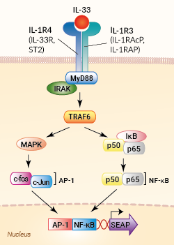
Signaling pathway in HEK-Blue™ IL-33 cells
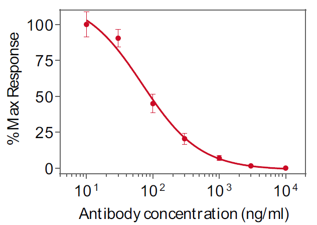
HEK-Blue™ IL-33 cells were engineered from the human embryonic kidney HEK293 cell line to detect bioactive interleukin-33 (IL-33) by monitoring the activation of the NF-κB and AP-1 pathways. In addition, these cells can be used for screening antibodies or small molecule inhibitors targeting the IL-33 pathway.
IL-33 is a member of the IL-1 family, a group of cytokines that play important roles in host defense, immune regulation and inflammation [1, 2].
Cell line description
HEK-Blue™ IL-33 cells were generated by stable transfection with the gene encoding the IL-1R4 chain (also known as IL-33R, or ST2) of the IL-33 receptor to obtain a fully active IL-33 signaling pathway. The other receptor subunit, IL-1R3 (aka IL-1RAcP), is naturally expressed in these cells. They were further transfected with an NF-κB/AP-1-inducible secreted embryonic alkaline phosphatase (SEAP) reporter. The binding of IL-33 to its receptor triggers a signaling cascade leading to NF-κB/AP-1 activation and the subsequent production of SEAP. This can be readily assessed in the supernatant using QUANTI-Blue™ Solution, a SEAP detection reagent.
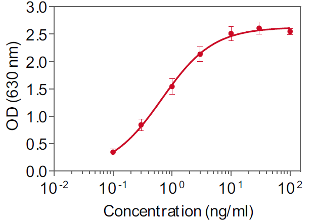
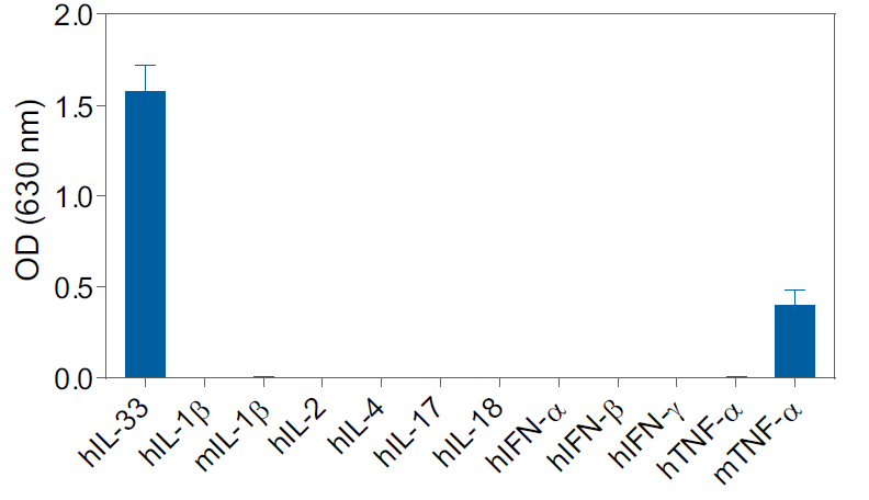
HEK-Blue™ IL-33 cells detect human IL-33 (see figures). Of note, they do not respond to human and murine IL-1β, nor to human TNF-α cells. However, these cells also respond, to a weaker extent, to murine TNF-α (see figures).
Key features
- Fully functional IL-33 signaling pathway
- Readily assessable NF-κB/AP-1-SEAP reporter activity
- Strong response to human IL-33
- No response to human (h) and murine (m) IL-1β, as well as hTNF-α
- Low response to mTNF-α
Applications
- Detection and quantification of human IL-33 activity
- Screening of anti-IL-33 or anti-IL33 receptor antibodies
- Screening of small molecule inhibitors of the IL-33 pathway
References:
1. Arend W. et al., 2008. IL-1, IL-18, and IL-33 families of cytokines. Immunol Rev. 223:20-38.
2. Mantovani, A., et al., 2019. Interleukin-1 and Related Cytokines in the Regulation of Inflammation and Immunity. Immunity. 50(4): p. 778-795.
Specifications
Antibiotic resistance: blasticidin, hygromycin B, Zeocin®
Growth medium: DMEM, 4.5 g/l glucose, 2 mM L-glutamine, 10% (v/v) heat-inactivated fetal bovine serum, 100 U/ml penicillin, 100 μg/ml streptomycin, 100 μg/ml Normocin™
Guaranteed mycoplasma-free
Specificity: Detects human IL-33
Detection range: 0.5 - 100 ng/ml
Back to the topContents
- 1 vial containing 3-7 x 10⁶ cells
- 2 x 1 ml of HEK-Blue™ Selection (250x concentrate)
- 1 ml of Normocin™ (50 mg/ml)
- 1 ml of QB reagent and 1 ml of QB buffer (sufficient to prepare 100 ml of QUANTI-Blue™ Solution, a SEAP detection reagent)
![]() Shipped on dry ice (Europe, USA, Canada and some areas in Asia)
Shipped on dry ice (Europe, USA, Canada and some areas in Asia)
Details
Interleukin-33 (IL-33; also known as IL-1F11, DVS22, NF-HEV)) is a member of the IL-1/Toll-like receptor cytokine superfamily, a group of cytokines that play important roles in host defense, immune regulation, and inflammation [1, 2].
IL-33 is constitutively expressed in lymphoid tissues, epithelial cells, fibroblasts, mucosal tissues, tumor cells, and vascular tissues [3]. It plays a central role in type 2 innate and adaptive immunity and inflammation, modulating Th2, ILC2 and M2 macrophage responses [2]. It is involved in responses to type 2 infections (e.g. parasites), tissue repair, as well as harmful allergic responses (e.g. asthma) [2].
IL-33 mediates its biological effects through the IL-1R4 receptor (also known as ST2, IL1RL1, IL-33R) and the IL-1R3 accessory protein (also known as IL-1RAcP or IL-1RAP) [2, 4, 5].
Upon ligand binding, IL-1Rs dimerize through their Toll/interleukin-1 resistance (TIR) domains to recruit the MyD88 adaptor protein, which then couples to IL-1R-associated kinases (IRAKs) and tumor necrosis factor receptor-associated factor 6 (TRAF6). This leads to the activation of key transcription factors, including NF-KB, AP-1, and IRFs [2, 4].
IL-33 can function both as a traditional cytokine and as a nuclear factor regulating gene transcription. Following pro-inflammatory stimulation, IL-33 can induce Th2-biased immune responses, such as the production of IL-4, IL-5 and IL-13 [6]. In addition, as IL-33 is constitutively expressed in endothelial and epithelial cells, it can act as an endogenous danger signal, or damage-associated molecular pattern (DAMP; also called alarmins), in response to tissue damage [7, 8].
IL-33 has emerged as a key regulatory cytokine in barrier tissues, making it an important target for inhibition therapy in autoimmune diseases such as inflammatory bowel disease (IBD), rhumatoid arthgritis (RA), asthma, and allergies.
1. Arend W. et al., 2008. IL-1, IL-18, and IL-33 families of cytokines. Immunol Rev. 223:20-38.
2. Mantovani, A., et al., 2019. Interleukin-1 and Related Cytokines in the Regulation of Inflammation and Immunity. Immunity. 50(4): p. 778-795.
3. Catalan-Dibene, J. et al., 2018. Interleukin 30 to Interleukin 40. J Interferon Cytokine Res. 38(10):423-439.
4. Teufel, L.U., et al., 2022. IL-1 family cytokines as drivers and inhibitors of trained immunity. Cytokine. 150: p. 155773.
5. Gaballa, J.M., et al., 2024. International nomenclature guidelines for the IL-1 family of cytokines and receptors. Nature Immunology. 25(4): p. 581-582.
6. Schiering C. et al., 2014. The alarmin IL-33 promotes regulatory T-cell function in the intestine. Nature. 513(7519):564-8.
7. Cayrol C. & Girard JP., 2014. IL-33: an alarmin cytokine with crucial roles in innate immunity, inflammation and allergy. Curr Opin Immunol. 31-7.
8. Oboki K. et al., 2011. IL-33 and airway inflammation. Allergy Asthma Immunol Res. 3(2): 81–88.


