IFN-α/β Reporter HEK 293 Cells
| Product | Unit size | Cat. code | Docs. | Qty. | Price | |
|---|---|---|---|---|---|---|
|
HEK-Blue™ IFN-α/β Cells Human HEK293 cells - Type I IFNs Reporter Cells |
Show product |
3-7 x 10e6 cells |
hkb-ifnabv2
|
|||
|
HEK-Blue™ IFN-α/β vial Additional cell vial |
Show product |
3-7 x 10e6 cells |
hkb-ifnabv2-av
|
![]() Cytokine offer: For each cytokine reporter cell line purchased, get a free vial of the matching cytokine.
Cytokine offer: For each cytokine reporter cell line purchased, get a free vial of the matching cytokine.
Human Type I IFNs Reporter Cells

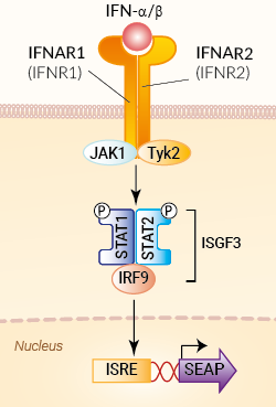
Signaling pathway in HEK-Blue™ IFN-α/β cells
InvivoGen also offers:
• Recombinant human IFN-α2b
• Anti-hIFNAR (Anifrolumab)
• Other IFN-α subtypes upon request
HEK-Blue™ IFN-α/β cells were engineered from the human embryonic kidney HEK 293 cell line to detect bioactive human type I interferons (e.g. IFN-α, IFN-β) by monitoring the activation of the JAK/ISGF3 (STAT1/STAT2/IRF9 complex) pathway. In addition, these cells can be used for screening antibodies or small molecule inhibitors targeting the IFN-α/β pathway.
IFN-α and IFN-β are important anti-viral cytokines that also have anti-proliferative and immunomodulatory functions [1, 2].
Cell line description
HEK-Blue™ IFN-α/β cells were generated by stable transfection with the genes encoding human STAT2 and IRF9 to obtain a fully active type I IFN signaling pathway. The other genes of the pathway (IFNAR1, IFNAR2, JAK1, TyK2, and STAT1) are naturally expressed by these cells. HEK-Blue™ IFN-α/β cells were also stably transfected with the secreted embryonic alkaline phosphatase (SEAP) reporter under the control of the ISG54 promoter. This promoter comprises IFN-stimulated response elements (ISRE) that are recognized by the ISGF3 complex. The binding of IFN-α or IFN-β to their receptor triggers a signaling cascade leading to the activation of ISGF3 and the subsequent production of SEAP. This can be readily assessed in the supernatant using QUANTI-Blue™ Solution, a SEAP detection reagent.
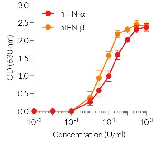
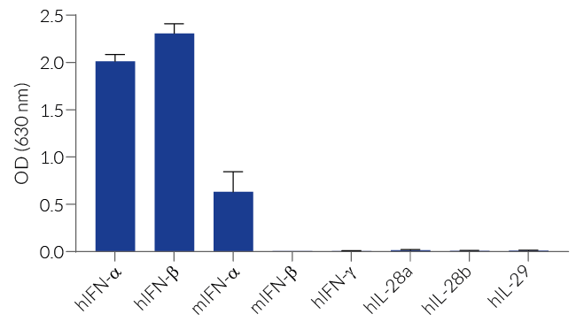
HEK-Blue™ IFN-α/β cells detect human (h) IFN-α and hIFN-β. At high concentrations, they might respond to mouse (m) IFN-α, but not mIFN-β (see figures). Of note, HEK-Blue™ IFN-α/β cells do not respond to human type II and type III IFNs (IFN-γ/λ) (see figures).
Key Features
- Fully functional human IFN-α/β signaling pathway
- Readily assessable ISGF3-inducible SEAP reporter activity
- Strong response to human (h) IFN-α and hIFN-β
- Poor to no response to murine (m) IFN-α and mIFN-β
- No response to IFN-γ and IFN-λ
Applications
- Detection of human IFN-α and IFN-β
- Quantification of IFN-α/β activity levels in biological samples, such as plasma or serum [3]
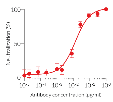
- Screening of anti-IFN-α/β and anti-IFNAR antibodies
- Screening of small molecule inhibitors of the IFN-α/β pathway
Note: A new clone is provided with an improved Type I IFN response. The cat code has been changed accordingly (hkb-ifnabv2).
References:
1. Schreiber G. 2017. The molecular basis for differential type I interferon signaling. J. Biol. Chem. 292:7285-94.
2. McNab F. et al., 2015. Type I interferons in infectious disease. Nat Rev Immunol. 15(2):87-103.
3. Gómez-Bañuelos E, et al., 2024. Uncoupling interferons and the interferon signature explain clinical and transcriptional subsets in SLE. medRxiv. 2023.08.28.23294734
Specifications
Antibiotic resistance: Blasticidin, Zeocin®
Growth medium: DMEM, 4.5 g/l glucose, 2 mM L-glutamine, 10% (v/v) heat-inactivated fetal bovine serum, 100 U/ml penicillin, 100 µg/ml streptomycin, 100 µg/ml Normocin®
Specificity: Detects human IFN-α and human IFN-β
Detection range:
- Detection range for human IFN-α: 1 - 103 U/ml
- Detection range for human IFN-β: 1 - 103 U/ml
Quality Control:
- SEAP reporter activity in response to human IFN-α and human IFN-β is validated using functional assays.
- The stability for 20 passages following thawing is confirmed.
- These cells are tested for mycoplasma contamination.
Contents
HEK-Blue™ IFN-α/β Cells (hkb-ifnabv2)
- 1 vial containing 3-7 x 10⁶ cells
- 1 ml of Blasticidin (10 mg/ml)
- 1 ml of Zeocin® (100 mg/ml)
- 1 ml Normocin® (50 mg/ml)
-
1 ml of QB reagent and 1 ml of QB buffer (sufficient to prepare 100 ml of QUANTI-Blue™ Solution, a SEAP detection reagent)
HEK-Blue™ IFN-α/β vial (hkb-ifnabv2-av)
-
1 vial containing 3-7 x 10⁶ cells
![]() Shipped on dry ice (Europe, USA, Canada and some areas in Asia)
Shipped on dry ice (Europe, USA, Canada and some areas in Asia)
Notification: Reference #hkb-ifnabv2-av can only be ordered together with reference #hkb-ifnabv2.
Back to the topDetails
Type I interferons, in particular interferon-alpha (IFN-α) and interferon beta (IFN-β), play a vital role in host resistance to viral infections [1, 2]. The type I IFN family is a multi-gene cytokine family that encodes 13 partially homologous IFN-α subtypes in humans (14 in mice), a single IFN-β, and several poorly defined single gene products (IFN-ɛ, IFN-τ, IFN-κ, IFN-ω, IFN-δ, and IFN-ζ) [1, 2]. IFN-α and IFN-β are the best-defined and most broadly expressed type I IFNs [2].
IFN-β and all of the IFN-α subtypes bind to a heterodimeric transmembrane receptor composed of the subunits IFNAR1 and IFNAR2 which are associated with the tyrosine kinases Tyk2 and Jak1 (Janus kinase 1) respectively. These kinases phosphorylate STAT1 and STAT2 which then dimerize and interact with IFN regulatory factor 9 (IRF9), leading to the formation of the ISGF3 complex. ISGF3 binds to IFN-stimulated response elements (ISRE) in the promoters of IFN-stimulated genes (ISG) to regulate their expression.
Despite their protective effects, studies have shown that aberrantly expression of the type I IFN system can elicit autoimmune disorders, such as interferonopathies and SLE (systemic lupus erythematosus). Recent evidence also implicates type I IFN-dependent signaling as a key inflammatory driver in non-autoimmune diseases [3].
References:
1. Schreiber G. 2017. The molecular basis for differential type I interferon signaling. J. Biol. Chem. 292:7285-94.
2. McNab F. et al., 2015. Type I interferons in infectious disease. Nat Rev Immunol. 15(2):87-103.
3. Crow MK, Ronnblom L. Type I interferons in host defence and inflammatory diseases. Lupus Sci Med. 2019 May 28;6(1):e000336.


