THP1-Null2 Cells
| Product | Unit size | Cat. code | Docs. | Qty. | Price | |
|---|---|---|---|---|---|---|
|
THP1-Null2 Cells Positive control cell line for inflammasome studies |
Show product |
3-7 x 10^6 cells |
thp-nullz
|
|
||
|
THP1-Null2 vial Additional cell vial |
Show product |
3-7 x 10e6 cells |
thp-nullz-av
|
Notification: Reference #thp-nullz-av can only be ordered together with reference #thp-nullz.
Positive control cell line for inflammasome studies
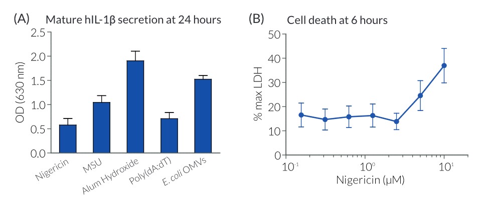
THP1-Null2 cells are derived from THP-1 human monocytic cells, the most commonly used model cell line for the study of inflammasome activation. Indeed, they express high levels of NLRP3, ASC, and pro-caspase-1 [1]. These cells produce IL-1β upon stimulation with canonical or non-canonical inflammasome inducers, such as Nigericin, MSU crystals, Alum Hydroxide, Poly(dA:dT), LPS, or E. coli outer membrane vesicles (OMVs). Moreover, they undergo pyroptotic cell death upon inflammasome activation.
THP1-Null2 cells are the positive control cell line for InvivoGen’s THP1-KO-ASC, THP1-KO-NLRP3, THP1-KO-CASP4, and THP1-KO-GSDMD cells.
These cells are resistant to Zeocin®.
For detecting and quantifying the release of mature human (h)IL-1β, InvivoGen provides HEK-Blue™ IL-1β sensor cells, which express an NF-κB-inducible SEAP reporter gene. QUANTI-Blue™ Solution allows rapid colorimetric detection and measure of SEAP activity by reading the optical density at 630-650 nm.
![]() Read our review on Inflammasome activation
Read our review on Inflammasome activation
References:
1. Schmid-Burgk, J.L et al., 2015. Caspase-4 mediates non-canonical activation of the NLRP3 inflammasome in human myeloid cells. Eur. J. Immunol. 45:2911.
Back to the topSpecifications
Antibiotic resistance: Zeocin®
Growth Medium: RPMI 1640, 2 mM L-glutamine, 25 mM HEPES, 10% heat-inactivated fetal bovine serum, 100 μg/ml Normocin™, Pen-Strep (100 U/ml-100 μg/ml)
Guaranteed mycoplasma-free
Quality control:
- The functionality of THP1-Null2 cells was tested using inflammasome inducers, such as Nigericin, MSU crystals, Alum Hydroxide, and E. coli OMVs.
Contents
- 3-7 x 10⁶ THP1-Null2 cells in a cryovial or shipping flask.
- 1 ml of Zeocin® (100 mg/ml). Store at 4 °C or at -20 °C.
- 1 ml of Normocin™ (50 mg/ml). Normocin™ is a formulation of three antibiotics active against mycoplasmas, bacteria, and fungi. Store at -20 °C.
IMPORTANT: If cells are shipped frozen (i.e. in a cryovial) and are not frozen upon arrival, contact InvivoGen immediately.
![]() Shipped on dry ice (Europe, USA, Canada and some areas in Asia)
Shipped on dry ice (Europe, USA, Canada and some areas in Asia)
Details
THP1-Null2 cells are suited for the study of inflammasome activation. The model of functional inflammasome formation is a two-step process of priming (step 1) followed by activation (step 2). Priming (i.e. with LPS) induces the production of pro-IL-1β, the immature form of IL-1β. Subsequent stimulation with inflammasome inducers, such as Nigericin or Alum Hydroxide, leads to caspase-1 activation and IL-1β maturation and secretion. Mature IL-1β can be detected by Western blot, ELISA, or a cell-based assay.
InvivoGen has developed a cellular assay to detect bioactive IL-1β using HEK-Blue™ IL-1β reporter cells. These cells feature the SEAP (secreted embryonic alkaline phosphatase) reporter gene under the control of an NF-kB-inducible promoter. They naturally express the IL-1β receptor (IL-1R), and all the proteins involved in the MyD88-dependent IL-1R signaling pathway that leads to NF-kB activation. Thus, upon IL-1β binding to IL-1R, a signaling cascade is initiated triggering NF-kB activation and the subsequent production of SEAP. Detection of SEAP in the supernatant of HEK-Blue™ IL-1β cells can be readily assessed using QUANTI-Blue™ Solution, a SEAP detection medium. QUANTI-Blue™ Solution turns blue in the presence of SEAP which can be easily quantified using a spectrophotometer.
Back to the top


