Pyroptosis & Necroptosis Cellular Reporter Assay
| Product | Unit size | Cat. code | Docs. | Qty. | Price | |
|---|---|---|---|---|---|---|
|
THP1-HMGB1-Lucia™ Cells Human THP-1 monocytes - HMGB1 release reporter cells |
Show product |
3-7 x 10e6 cells |
thp-gb1lc
|
|
||
|
THP1-HMGB1-Lucia™ vial Additional cell vial |
Show product |
3-7 x 10e6 cells |
thp-gb1lc-av
|
Notification: Reference #thp-gb1lc-av can only be ordered together with reference #thp-gb1lc.
HMGB1-Lucia reporter assay for pyroptosis and necroptosis monitoring

Signaling in THP1-HMGB1-Lucia™ cells
High mobility group box 1 (HMGB1) plays a critical role in the stress response not only inside the cell as a DNA chaperone and cell death regulator but also outside the cell as a prototypical alarmin. Indeed, during non-apoptotic programmed or regulated cell death, such as pyroptosis and necroptosis, HMGB1 is released along with other pro-inflammatory molecules [1-3].
![]() More details about Pyroptosis and necroptosis
More details about Pyroptosis and necroptosis
THP1-HMGB1-Lucia™ cells are designed to monitor necroptosis and inflammasome-mediated pyroptosis, two forms of necrotic cell death characterized by the release of the alarmin HMGB1 upon cell membrane rupture [1-3]. These cells are derived from the THP-1 human monocytic cell line and stably express a HMGB1::Lucia-luciferase fusion protein. They respond to commonly used inflammasome inducers and necroptosis cocktail inducers. The release of the HMGB1::Lucia protein in the extracellular milieu upon pyroptosis or necroptosis can be readily measured using QUANTI-Luc™ 4 Lucia/Gaussia, a Lucia and Gaussia luciferase detection reagent.
Key features
- Pyroptosis and necroptosis reporter cells
- Readily assessable HMGB1::Lucia reporter activity
- Guaranteed mycoplasma-free
Levels of IL-1β can be measured by Western blot, ELISA, or using InvivoGen’s HEK-Blue™ IL-1β cellular assay. Alternatively, secreted IL-18 can be detected using InvivoGen’s HEK-Blue™ IL-18 cellular assay.
![]() Download our Practical guide on Inflammasomes
Download our Practical guide on Inflammasomes
References:
1. Broz P. and Dixit V.M., 2016. Inflammasomes: mechanism of assembly, regulation and signalling. Nat Rev Immunol. 16:407-20.
2. Kovacs S.B. and Miao E.A., 2017. Gasdermins: effectors of pyroptosis. Trends Cell Biol. 27:673-84.
3. Grootjans S. et al., 2017. Initiation and execution mechanisms of necroptosis: an overview. Cell Death Differ. 24:1184-95.
Specifications
Antibiotic resistance: Zeocin®
Growth medium: RPMI 1640, 2 mM L-glutamine, 25 mM HEPES, 10% heat-inactivated fetal bovine serum (FBS), 100 U/ml penicillin, 100 μg/ml streptomycin,100 μg/ml Normocin™
Quality control:
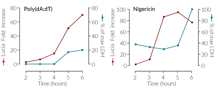
- The functionality of THP1-HMGB1-Lucia™ cells was tested using inflammasome inducers, such as Nigericin.
- The stability of this cell line for 20 passages following thawing has been verified.
- THP1-HMGB1-Lucia™ cells are guaranteed mycoplasma-free.
Contents
- 3-7 x 10⁶ THP1-HMGB1-Lucia™ cells in a cryovial or shipping flask
- 1 ml Normocin™ (50 mg/ml). Normocin™ is a formulation of three antibiotics active against mycoplasmas, bacteria, and fungi.
- 100 µl Zeocin® (100 mg/ml).
- 1 tube of QUANTI-Luc™ 4 Reagent, a Lucia luciferase detection reagent (sufficient to prepare 25 ml)
![]() Shipped on dry ice (Europe, USA, Canada, and some areas in Asia)
Shipped on dry ice (Europe, USA, Canada, and some areas in Asia)
Details
Pyroptosis is a consequence of inflammasome activation.
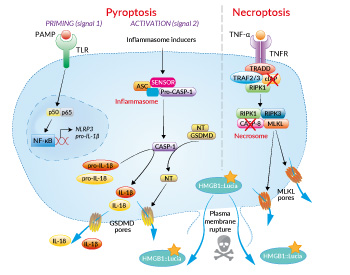
Typically, inflammasome activation is a two-step process. A first signal (‘priming’), provided by microbial molecules such as lipopolysaccharide (LPS), induces NF-κB-dependent expression of pro-IL1β. The second signal, provided by structurally unrelated microbial molecules (e.g. Nigericin toxin) or danger signals, triggers inflammasome multimerization. This leads to caspase-1 self-activation, proteolytic maturation of IL-1β and IL-18, and cleavage of Gasdermin D (GSDMD). Subsequent GSDMD pore formation at the cell membrane elicits a rapid cell death associated with the release of IL-1β, IL-18, and HMGB1 [1,2].
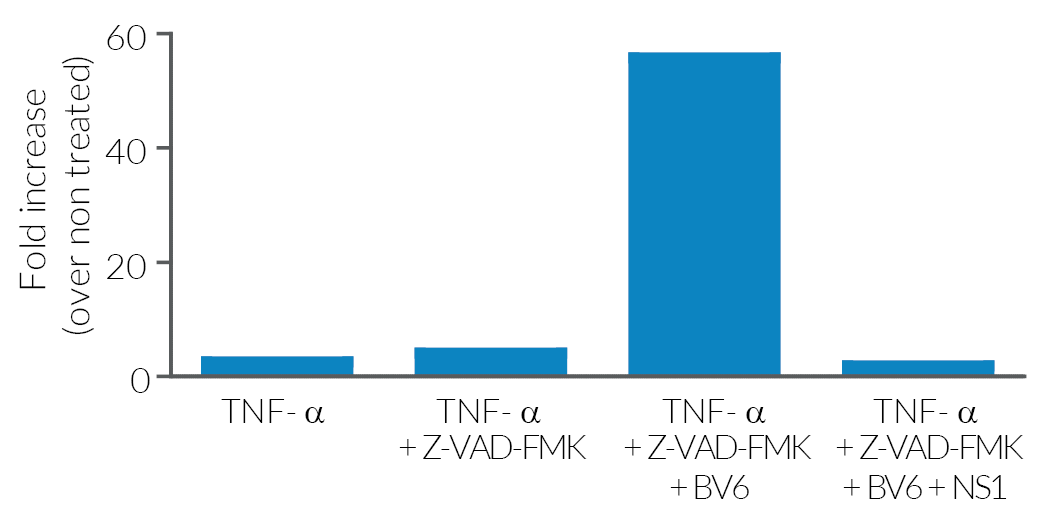
Necroptosis is mainly implicated in conditions of disease or infection and is triggered by the formation of a necrosome. The necrosome is induced upon Tumor Necrosis Factor (TNF) receptor or PRR activation in absence of caspase-8 and cIAP (cellular inhibitor of apoptosis) activity. The necrosome contains receptor-interacting serine/threonine-protein kinases RIPK1 and RIPK3, and the mixed lineage kinase-like MLKL. Phosphorylated MLKL relocalizes at the plasma membrane where it drives cell lysis and release of HMGB1, most probably through pore formation [3].
1. Broz P. and Dixit V.M., 2016. Inflammasomes: mechanism of assembly, regulation and signalling. Nat Rev Immunol. 16:407-20.
2. Kovacs S.B. and Miao E.A., 2017. Gasdermins: effectors of pyroptosis. Trends Cell Biol. 27:673-84.
3. Grootjans S. et al., 2017. Initiation and execution mechanisms of necroptosis: an overview. Cell Death Differ. 24:1184-95.


