TBK1 KO Dual Reporter THP1 Cells
| Product | Unit size | Cat. code | Docs. | Qty. | Price | |
|---|---|---|---|---|---|---|
|
THP1-Dual™ KO-TBK1 Cells Human THP-1 Monocytes - TBK1 knockout NF-κB-SEAP and IRF-Lucia Reporter Cells |
Show product |
3-7 x 10e6 cells |
thpd-kotbk
|
|
||
|
THP1-Dual™ KO-TBK1 vial Additional cell vial |
Show product |
3-7 x 10e6 cells |
thpd-kotbk-av
|
Notification: Reference #thpd-kotbk-av can only be ordered together with reference #thpd-kotbk.
TBK1 knockout dual reporter monocytes

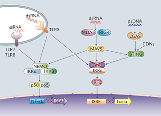
Nucleic acid sensing signaling pathways in THP1-Dual™ cells
THP1-Dual™ KO-TBK1 cells were generated from the THP1-Dual™ cell line, which is derived from the human THP-1 monocytes, through the stable knockout of the TBK1 gene. TBK1 (TANK-binding kinase 1) functions as a key node protein in several cell signaling pathways, including innate immune responses elicited upon pattern recognition receptor (PRR) activation. Upon ligand (e.g. cytoplasmic nucleic acids) binding, these receptors trigger the production of interferons (IFNs) through the activation of the TBK1/IKKε-IRF3/IRF7 (interferon regulatory factors 3 and 7) pathway [1]. Activated PRRs also trigger the production of pro-inflammatory cytokines through activation of the NF-κB pathway [1].
THP1-Dual™ KO-TBK1 and THP1-Dual™ cells feature two reporter genes allowing the simultaneous study of the IRF pathway, by monitoring the activity of an inducible secreted Lucia luciferase, and the NF-κB pathway by monitoring the activity of an inducible SEAP (secreted embryonic alkaline phosphatase). Lucia luciferase and SEAP activities are readily assessable in the supernatant using QUANTI-Luc™ 4 Lucia/Gaussia and QUANTI-Blue™ Solution detection reagents, respectively.
As expected, the responses of THP1-Dual™ KO-TBK1 cells to the cyclic dinucleotide 2'3'-cGAMP (a STING agonist) and to lipopolysaccharide (a TLR4 agonist) are impaired. However, differential responses (either no effect, decrease or increase) are observed when using RNA or DNA agonists coupled with transfection reagents. THP1-Dual™ KO-TBK1 cells retain the ability to respond to cytokines such as type I IFNs and TNF-α. These cells are resistant to Blasticidin and Zeocin™.
Key Features:
- Verified biallelic knockout of the TBK1 gene by PCR, DNA sequencing, Western blot, and functional assays
- Readily assessable Lucia luciferase and SEAP reporter activity
Applications:
- Defining the role of TBK1 in PRR-induced signaling, or other cell signaling pathways (e.g. cell growth)
- Highlighting possible overlapping PRR activation or regulatory mechanisms (see 'Details' tab)
References:
1. Iurescia S. et al., 2018. Nucleic acid sensing machinery: targeting innate immune system for cancer therapy. Recent Pat. Anticancer Drug Discov. 13: 2-17
Back to the topSpecifications
Antibiotic resistance: Blasticidin and Zeocin®
Growth medium: RPMI 1640, 2 mM L-glutamine, 25 mM HEPES, 10% (v/v) fetal bovine serum (FBS), 100 U/ml penicillin, 100 µg/ml streptomycin, 100 µg/ml Normocin™
Quality Control:
- Biallelic TBK1 knockout has been verified by PCR, DNA sequencing, western blot, and functional assays.
- The stability for 20 passages, following thawing, has been verified.
- These cells are guaranteed mycoplasma-free.
Contents
- 1 vial of THP1-Dual™ KO-TBK1 cells (3-7 x 10⁶ cells) in freezing medium
- 1 ml of Normocin™ (50 mg/ml). Normocin™ is a formulation of three antibiotics active against mycoplasmas, bacteria, and fungi.
- 1 ml of Zeocin® (100 mg/ml)
- 1 ml of Blasticidin (10 mg/ml)
- 1 tube of QUANTI-Luc™ 4 Reagent, a Lucia luciferase detection reagent (sufficient to prepare 25 ml)
- 1 ml of QB reagent and 1 ml of QB buffer (sufficient to prepare 100 ml of QUANTI-Blue™ Solution, a SEAP detection reagent)
![]() Shipped on dry ice (Europe, USA, Canada, and some areas in Asia)
Shipped on dry ice (Europe, USA, Canada, and some areas in Asia)
Details
THP1-Dual™ KO-TBK1 cells have been functionally tested using various sources of nucleic acids (see the validation data document).
As expected, IRF and NF-κB responses are severely impaired when the THP1-Dual™ KO-TBK1 cells are incubated with STING agonists, such as 2’3’-cGAMP, which do not require transfection to access the cytosol. However, differential responses are observed when using RNA agonists with transfection reagents. The weak RIG-I agonist 5’ppp-dsRNA loses its ability to induce an IRF response in KO-TBK1 cells when complexed with LyoVec™ or LTX. On the contrary, the IRF response is unexpectedly increased when using the highly potent RIG-I agonist 3p-hpRNA complexed to LyoVec™ or LTX. Surprisingly, with either agonist, the NF-κB response is barely affected, if not slightly increased. These data suggest that the use of different agonists and transfection reagents could highlight overlapping RNA-sensing or regulatory mechanisms [1, 2].
References:
1. Perry AK. et al., 2004. Differential requirement for TANK-binding kinase-1 in type I interferon responses to Toll-like receptor activation and viral infection. J. Exp. Med. 199:1651.
2. Deng W. et al., 2008. Negative regulation of virus-triggered IFN-B signaling pathway by alternative splicing of TBK1. J. Biol. Chem. 283:35590.


